Californian Researchers 3D Print functioning blood vessels

CALIFORNIAN RESEARCHERS 3D PRINT FUNCTIONING BLOOD VESSELS
Researchers from the University of California, San Diego have successfully 3D printed a framework of functional blood vessels. Blood vessel networks are important in transporting blood, nutrients and waste around the human body.
The research team employed a 3D bioprinting process involving hydrogel and endothelial cells. Endothelial are the form of cells that make up the inner lining of blood vessels.
Leading the research was Shaochen Chen, who explains the motivation of the project,
ALMOST ALL TISSUES AND ORGANS NEED BLOOD VESSELS TO SURVIVE AND WORK PROPERLY. THIS IS A BIG BOTTLENECK IN MAKING ORGAN TRANSPLANTS, WHICH ARE IN HIGH DEMAND BUT IN SHORT SUPPLY. 3D BIOPRINTING ORGANS CAN HELP BRIDGE THIS GAP, AND OUR LAB HAS TAKEN A BIG STEP TOWARD THAT GOAL.
Vessel bioprinting process
This technique is novel because the laboratory has successfully bioprinted vessels that can be incorporated into the body’s existing circulatory network. These structures can branch out into smaller vessels to fully integrate into the body’s cardiovascular system. Whereas other research has focused on printing a single vessel, such as researchers in China recently implementing a 3D printed blood vessel into macaque monkeys.
To create the 3D printed vessels, the Californian researchers implemented a Digital Light Processing method. Using hydrogel and encapsulated cells to mimic the “native vascular cell composition.”
Nanoengineering professor Shaochen Chen, who also leads the Nanobiomaterials, Bioprinting, and Tissue Engineering Lab at UC San Diego has received acclaim for previous 3D printing research. His laboratory successfully 3D printed micro-fish that could monitor the workings of a human body.
Testing on mice
The research paper explains how the vessels functioned normally once implanted as part of an existing structure. When the 3D printed vessels were implanted in mice in order to test their functionality researchers observed that two weeks after grafting, the vessels had correctly merged with the existing framework and were circulating blood.
While successful, the vessels are currently unable to transfer nutrients and waste. As Chen says, “we still have a lot of work to do to improve these materials.” In the meantime, Chen’s team plan to to create specific tissues using cells extracted from a patient. This would ensure the vessels are accepted by the host when implanted.

Digital model showing the structure of the blood vessels. Photo Erik Jepsen/UC San Diego Publications.
Future of 3D printed vessels
Following this initial success, Chen and his team aim to begin clinical trials, however he says, “It will take at least several years before we reach that goal.”
The research paper concludes that,
THIS PLATFORM CAN BE FURTHER EXTENDED TO ENGINEER OTHER TISSUES THAT FEATURE COMPLEX MICROARCHITECTURES, SUCH AS LIVER, HEART AND NERVE TISSUES.
The full paper ‘Direct 3D bioprinting of prevascularized tissue constructs with complex microarchitecture’ is published in the academic journal Biomaterials.
For the latest news on 3D bioprinting, follow us on twitter and instagram.
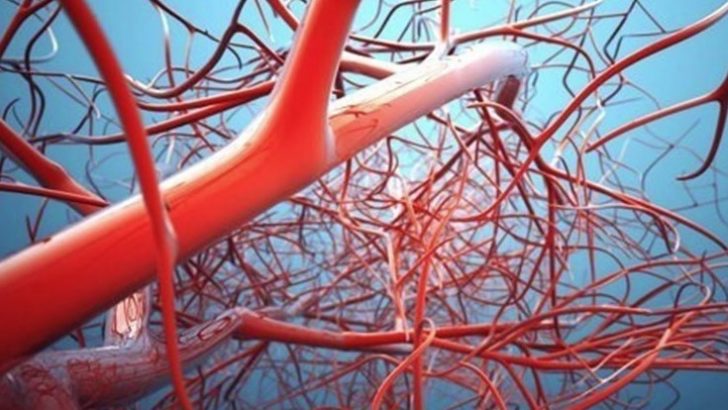
Featured image shows blood vessels in the brain in a piece from the Wellcome Collection, London, ‘Brains: The mind as matter’ exhibition. Photo via Wellcome Collection.
Search: 3dprintingindustry


